Back Bones Diagram - Diagram Of Vertebral Column Showing Different Parts And Regions Of The Download Scientific Diagram : The notochord present in the embryonic stage is replaced by the vertebral column.
byAdmin•
0
Back Bones Diagram - Diagram Of Vertebral Column Showing Different Parts And Regions Of The Download Scientific Diagram : The notochord present in the embryonic stage is replaced by the vertebral column.. Can you feel the bumps of your vertebrae along your back? Individual anatomical structures include 2: Anatomynote.com found anatomy of back muscles diagram from plenty of anatomical pictures on the internet. See sacrum (sacral region) the sacrum is connected to part of the pelvis (the iliac bones) by the sacroiliac joints. Its appearance is different from the other spinal vertebrae.
The temporal bone consists of a pair of bones that help make up the skull. A tough, springy disc of cartilage sits between the vertebrae of your spine. In the back and elsewhere in the body, tendons attach muscles to bones. The spine or backbone consists of 26 small bones or vertebrae. Individual anatomical structures include 2:
Basic Spinal Anatomy Welcome Back Clinic Mri And Pain Management Centre from www.welcomebackclinic.com Exercises can strengthen the core muscles that support the spine and. The bones of the appendicular skeleton provide support and flexibility at the. The first seven bones (vertebrae) of your spine form your neck. The bones of the pelvis and lower back work together to support the body's weight, anchor the abdominal and hip muscles, and protect the delicate vital organs of the vertebral and abdominopelvic cavities. The column can be divided into five different regions, with each region characterised by a different vertebral structure. The trapezius or trapezoid muscles are two paired muscles that extend from the base of the thoracic vertebrae in the spine to the occipital bone and run out to the spine of the scapula. Lower back bones diagram : Bones of the pelvis and lower back.
They help support particular bones and make them move.
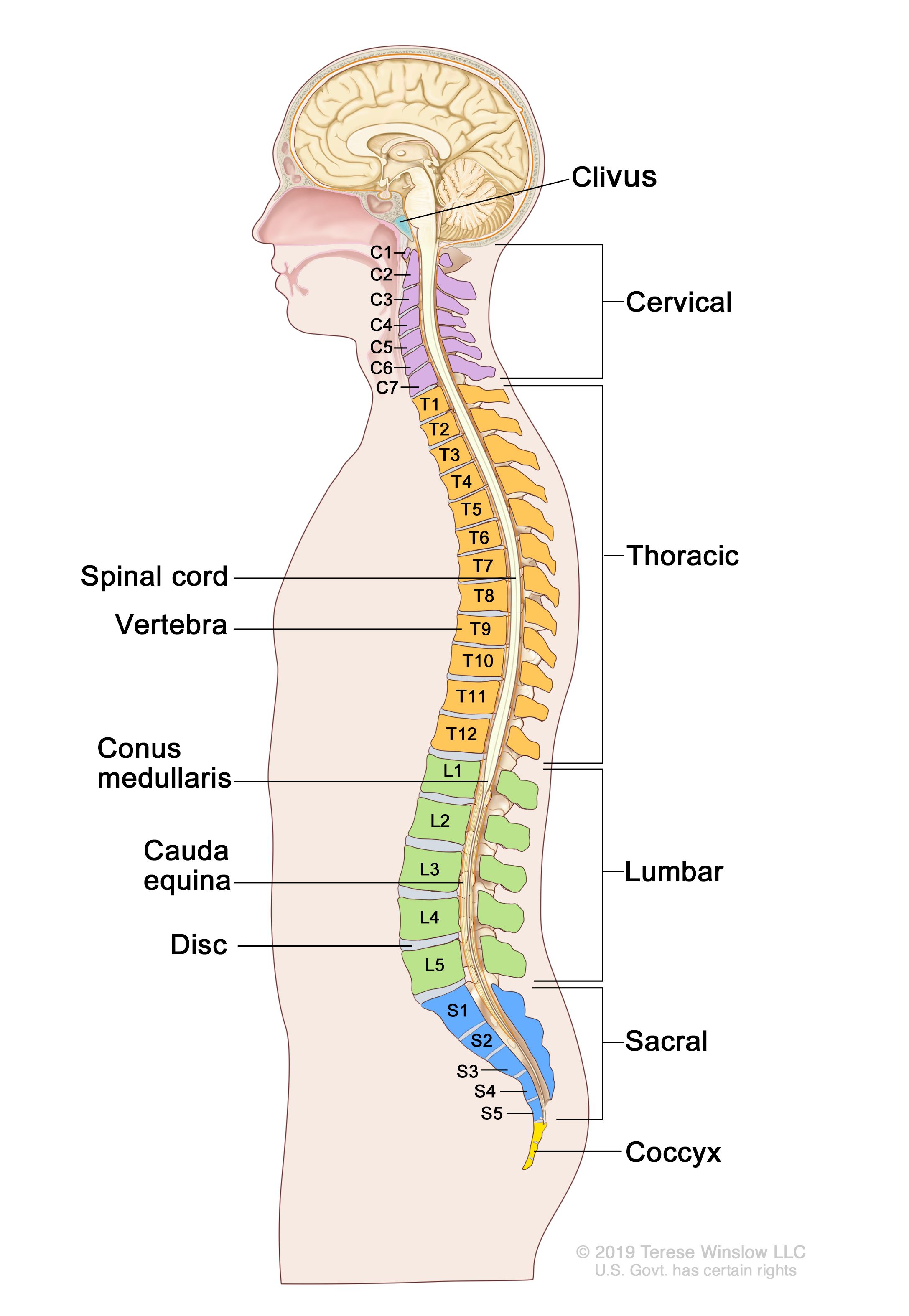
Bones of the pelvis and lower back. Studying a spine diagram is one way to better understand many of the individual components of the back bone and how they might relate to a symptomatic back, neck or sciatica pain condition. Skeleton back bones diagram / human skeleton anatomy vintage 1940s high res digital image / in this assignment, students color the various parts of the skeletal system and then answer some follow up teach your students the names of the bones in the human body with the help of this illustrated human skeleton diagram. They help support particular bones and make them move. The notochord present in the embryonic stage is replaced by the vertebral column. We think this is the most useful anatomy picture that you need. Diagram of a human female skeleton, back view. Each lumbar spinal level is numbered from top to bottom—l1 through l5, or l6. This bone is shaped like a triangle that fits between the two halves of the pelvis, connecting the spine to the lower half of the body. This vertebra supports the skull. Spinal vertebrae bone spine vertebra toracica spinal cord spine structure back diagram spine sections spinal cord vertebrae spinal structure health diagram. The occiput (co), also known as the occipital bone, is a flat bone that forms the back of the head. The vertebral column of the lower back includes the five lumbar vertebrae, the sacrum, and the coccyx.
In the back and elsewhere in the body, tendons attach muscles to bones. Back bones diagram / spencer system 1936. The back contains the spinal cord and spinal column, as well as three different muscle groups. Back of skull (occipital bone) fused vertebrae (5) (sacrum) hand bones (metacarpals) finger bones (phalanges) heel bone (calcaneus) skull (cranium) backbone The lumbar spine connects to the thoracic spine above and the hips below.
Anatomy Of The Back Spine And Back Muscles Kenhub from thumbor.kenhub.com Human backbone diagram, bone, human backbone diagram. 12 photos of the human back bone chart. The lumbar spine connects to the thoracic spine above and the hips below. The vast difference in height and limb length between birth and adulthood are mainly the result of endochondral ossification in the. See sacrum (sacral region) the sacrum is connected to part of the pelvis (the iliac bones) by the sacroiliac joints. The occiput (co), also known as the occipital bone, is a flat bone that forms the back of the head. This process continues until the end of puberty, when the growth plate stops growing and the bones fuse permanently into a single bone. Fishbone diagrams, aka ishikawa diagrams are used across various industries to analyze causes and their effect.
Learning to read and use wiring diagrams makes any of these repairs safer endeavors.
Diagram of a human female skeleton, back view. Lateral labeled diagram of the human vertebral spinal column showing vertebrae numbering order and the 5 different regions of the spine. The column can be divided into five different regions, with each region characterised by a different vertebral structure. Muscle or tendon injuries can occur anywhere in the body. We hope this picture anatomy of back muscles diagram can help you study and research. Its appearance is different from the other spinal vertebrae. The bones of the appendicular skeleton provide support and flexibility at the. Each lumbar spinal level is numbered from top to bottom—l1 through l5, or l6. The spine supports your body and helps you walk, twist and move. Human backbone diagram, bone, human backbone diagram. The spine diagram shown below, consists of many bones or vertebrae,soft discs,the spinal cord, and spinal nerves. Vertebrae are the structural constituents of the spine.there are 33 vertebrae in total; Lower limbs (60 bones, 30 each side).
Can you feel the bumps of your vertebrae along your back? A tough, springy disc of cartilage sits between the vertebrae of your spine. Each lumbar spinal level is numbered from top to bottom—l1 through l5, or l6. The atlas is the topmost vertebra, and along with c2, forms the joint connecting the skull and spine. The lower back is also associated with feeling unsupported (not backed up) by a family member, partner, friend, teacher, colleague, or employer.
Definition Of Backbone Nci Dictionary Of Cancer Terms National Cancer Institute from nci-media.cancer.gov The vast difference in height and limb length between birth and adulthood are mainly the result of endochondral ossification in the. The trapezius or trapezoid muscles are two paired muscles that extend from the base of the thoracic vertebrae in the spine to the occipital bone and run out to the spine of the scapula. A tough, springy disc of cartilage sits between the vertebrae of your spine. Each typical vertebra consists of a body, an arch and three processes that stem from. Bones of the pelvis and lower back. Fishbone diagrams, aka ishikawa diagrams are used across various industries to analyze causes and their effect. Seven cervical vertebrae in the neck, twelve thoracic vertebrae in the torso and five lumbar vertebrae in the lower back. The bones of the pelvis and lower back work together to support the body's weight, anchor the abdominal and hip muscles, and protect the delicate vital organs of the vertebral and abdominopelvic cavities.
Its appearance is different from the other spinal vertebrae.
At the back of each bone in the spine (vertebra) are bony points called processes, which muscles attach to. Vertebrae are the structural constituents of the spine.there are 33 vertebrae in total; The disks that cushion vertebrae may compress with age or injury, leading to a herniated disk. It is also known as the vertebral column. See sacrum (sacral region) the sacrum is connected to part of the pelvis (the iliac bones) by the sacroiliac joints. The notochord present in the embryonic stage is replaced by the vertebral column. A tough, springy disc of cartilage sits between the vertebrae of your spine. It also covers some common conditions and injuries that can affect the back. Its appearance is different from the other spinal vertebrae. The vertebral column of the lower back includes the five lumbar vertebrae, the sacrum, and the coccyx. The lower back is also associated with feeling unsupported (not backed up) by a family member, partner, friend, teacher, colleague, or employer. These bones are connected at the back with specialized joints. 12 photos of the human back bone chart.